Orthopedic diagnostics play a vital role in understanding and treating musculoskeletal conditions. Whether you’re dealing with an injury, chronic pain, or a degenerative condition, accurate diagnostics are essential for effective treatment and recovery. Premier Orthopaedics & Sports Medicine, P.C. specializes in utilizing advanced diagnostic tools such as X-rays, MRI, and CT scans to provide a comprehensive view of your musculoskeletal health. Our expert team in Bloomfield, Englewood, and Union City, NJ offers precise diagnosis and personalized care tailored to each patient’s unique needs.
How Premier Orthopaedics & Sports Medicine, P.C. Can Help You
Premier Orthopaedics & Sports Medicine, P.C.’s commitment to providing exceptional care begins with an accurate diagnosis. Our state-of-the-art imaging technologies allow us to pinpoint the root cause of your symptoms, enabling us to develop a targeted treatment plan. Whether you are experiencing joint pain, a sports-related injury, or any other orthopedic condition, our specialists are here to help.
- Comprehensive Evaluations: We conduct thorough evaluations to understand your condition fully. Our advanced imaging techniques help us visualize the affected areas and assess the extent of the damage, providing a clear picture of your orthopedic health.
- Personalized Treatment Plans: Once we understand your condition, we work with you to develop a customized treatment plan. Our approach ensures you receive the most effective treatment tailored to your needs.
- Experienced Team: Our orthopedic specialists have extensive experience in diagnosing and treating a wide range of musculoskeletal conditions. We are dedicated to staying at the forefront of medical advancements to provide the best care possible.
- Convenient Locations: With offices in Bloomfield, Englewood, and Union City, NJ, we offer easy access to high-quality orthopedic care. Our facilities are equipped with cutting-edge technology to ensure accurate and efficient diagnostics.
Advanced Diagnostic Tools: X-Rays, MRI, and CT Scans
X-Rays
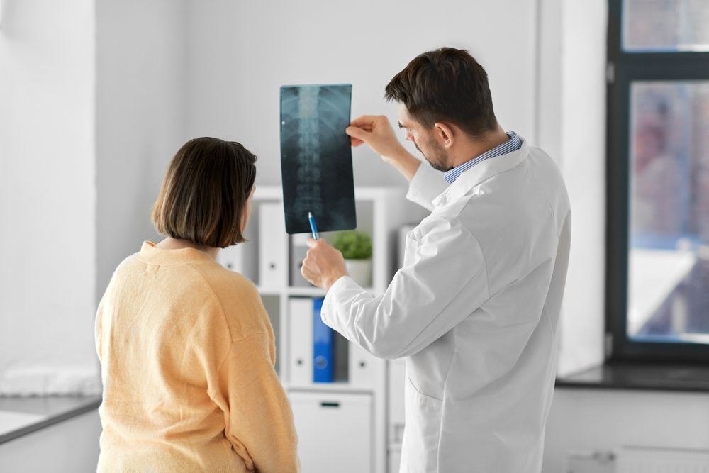
X-rays are one of the most common imaging techniques used in orthopedics. They are particularly useful for examining bones and joints.
- Bone Fractures: X-rays can quickly reveal fractures, breaks, and other bone abnormalities, helping us determine the best course of action for treatment.
- Joint Dislocation: X-rays provide a clear view of joint alignment, allowing us to diagnose dislocations and guide proper realignment.
- Arthritis: We use X-rays to assess joint space narrowing, bone spurs, and other signs of arthritis, enabling us to track disease progression and adjust treatment plans accordingly.
MRI (Magnetic Resonance Imaging)
MRI is a powerful imaging technique that provides detailed images of soft tissues, including muscles, ligaments, and cartilage.
- Soft Tissue Injuries: MRI is invaluable for diagnosing tears and injuries in muscles, ligaments, and tendons, offering a comprehensive view of soft tissue health.
- Spinal Disorders: MRI allows us to examine the spinal cord, discs, and nerves, helping us diagnose herniated discs and spinal stenosis.
- Tumors: With MRI, we can detect and evaluate tumors in bones and soft tissues, providing critical information for treatment planning.
CT Scans (Computed Tomography)
CT scans combine X-ray images from multiple angles to create cross-sectional body views, offering a more detailed picture than standard X-rays.
- Complex Fractures: CT scans are excellent for evaluating complex fractures. They provide detailed images of bone structure and help us plan surgical interventions if needed.
- Joint Problems: CT scans can visualize joint structures in detail, aiding in diagnosing conditions such as osteoarthritis and rheumatoid arthritis.
- Bone Tumors: CT scans help us assess bone tumors’ size, shape, and location, informing treatment decisions and monitoring response to therapy.
How Diagnostic Imaging Works
Diagnostic imaging is critical to modern orthopedic care, providing essential insights into various musculoskeletal conditions. Each imaging modality offers unique insights, and choosing the right one depends on the specific condition and diagnostic needs. X-rays are quick and painless procedures involving exposing a part of the body to a small dose of ionizing radiation. This radiation passes through the body and is captured on film or a digital detector, creating an image that shows different structures in varying shades of black and white. Dense materials like bone absorb more radiation and appear white, while softer tissues appear darker, making X-rays particularly useful for identifying bone fractures and joint dislocations.
MRI, on the other hand, uses powerful magnets and radio waves to generate detailed images of the body’s soft tissues. Unlike X-rays, MRI does not involve radiation, making it a safer option for imaging soft tissue structures. During an MRI, the patient lies inside a large magnet, and radio waves excite hydrogen atoms in the body, producing signals that are converted into images. This process results in a comprehensive view of muscles, tendons, ligaments, and other soft tissues, making MRI invaluable for diagnosing soft tissue injuries and spinal disorders.
CT scans combine X-ray technology and computer processing to create cross-sectional images of the body. In a CT scan, the patient lies on a table that moves through a doughnut-shaped scanner, while the X-ray tube rotates around the patient, capturing images from different angles. These images are then compiled to produce detailed cross-sectional views, allowing for a thorough examination of bone and soft tissue structures. CT scans are particularly beneficial for evaluating complex fractures and visualizing detailed joint structures, aiding in diagnosing various orthopedic conditions.
Frequently Asked Questions (FAQs)
1. How do I know which diagnostic imaging test is right for me?
The choice of imaging test depends on your symptoms, medical history, and the specific area of concern. Our orthopedic specialists will evaluate your condition and recommend the most appropriate imaging modality to ensure accurate diagnosis and treatment planning.
2. Are X-rays, MRIs, and CT scans safe?
Yes, these imaging techniques are generally safe when performed by trained professionals. X-rays and CT scans use low-dose radiation, while MRI does not involve radiation. Our team takes every precaution to minimize exposure and ensure your safety during imaging.
3. How should I prepare for an imaging test?
Preparation varies depending on the type of test. Little to no preparation is needed for X-rays. For MRIs, you may need to remove metal objects and inform your doctor of any implants. For CT scans, you might be asked to avoid eating or drinking for a few hours before the procedure. Our staff will provide detailed instructions based on the specific test.
Your Partner in Orthopedic Health
At Premier Orthopaedics & Sports Medicine, P.C., we are dedicated to providing the highest quality care and support to help you achieve optimal musculoskeletal health. Our advanced diagnostic imaging tools, including X-rays, MRI, and CT scans, allow us to accurately diagnose various orthopedic conditions, ensuring you receive the most effective treatment for your needs.
Our team of experienced specialists is committed to guiding you through every step of your orthopedic journey, from diagnosis to treatment and recovery. With our convenient locations in Bloomfield, Englewood, and Union City, NJ, we are here to provide exceptional care close to home. Schedule an appointment today and take the first step toward a healthier, more active life.
Sources:
- Reuben, D. B., & Siu, A. L. (1990). An Objective Measure of Physical Function of Elderly Outpatients: The Physical Performance Test. Journal of the American Geriatrics Society.
- Deyo, R. A., & Weinstein, J. N. (2001). Low Back Pain. New England Journal of Medicine.


